The geometry of generalization
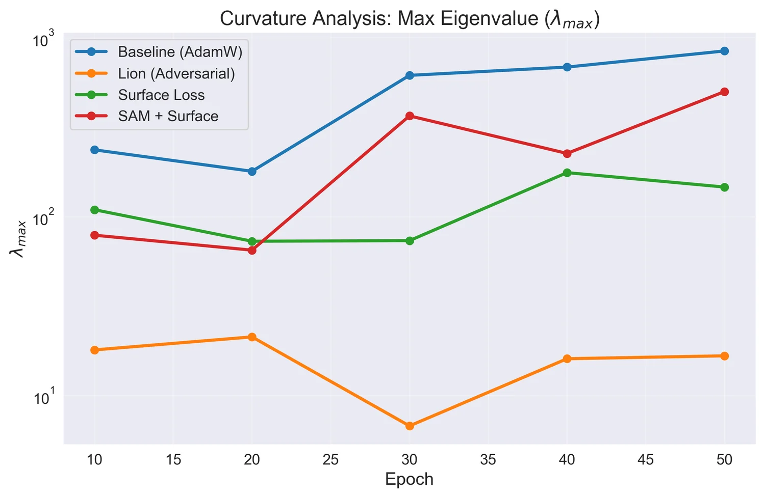
Four training runs on paediatric brain tumor MRI, held identical except for the loss and the optimiser, then read through the curvature of the loss surface rather than the leaderboard.
Medical imaging · implicit neural representations · deformable registration
Anatomy as a continuous function, not a grid of numbers.
I am finishing an M.S. in Computer Vision at the University of Central Florida, where I work on how to represent the deformation between two human bodies. My first author paper, QueryPhi, replaces the fixed voxel lattice that almost every method uses to store that deformation with a field you can evaluate at any coordinate.
It reaches the highest mean Dice of all ten methods on the IXI brain benchmark using a fraction of the parameters, keeps the warp near fold free, and carries the best boundary accuracy to a cohort it never trained on.
What I work with
The question
Given two scans of different people, deformable registration finds the smooth, invertible map that carries one onto the other. What that map has to do in order to succeed is precisely the difference between those two bodies. Atlases, morphometry, longitudinal studies and label transfer all rest on it.
The convention is to store that map as a displacement vector at every voxel of a fixed grid. QueryPhi stores it as a function instead, so the deformation has a value at every coordinate rather than only at the grid nodes, and its derivatives come out analytically.

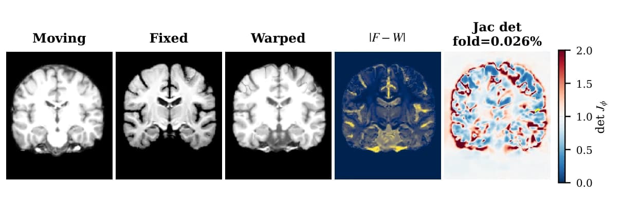
Two different people, and the smooth invertible map that carries one onto the other. What the map has to do in order to succeed is the difference between them.
NeurIPS 2026 · under review · first author
A registration model that stores the deformation as a function you can evaluate anywhere, rather than as numbers parked on a fixed voxel grid.
0.7726
IXI atlas Dice
first of ten, 95% CI 0.769 to 0.776
0.7534
Nearest method
TransMorph-bspl, p = 1.5e-8
5.51M
Parameters
against 46.8M for the transformers
0.0001%
Folded voxels
near fold free, empirical
1.502 mm
OASIS zero shot
best HD95 of nine methods
1.77
Average rank
Friedman and Nemenyi, ten methods
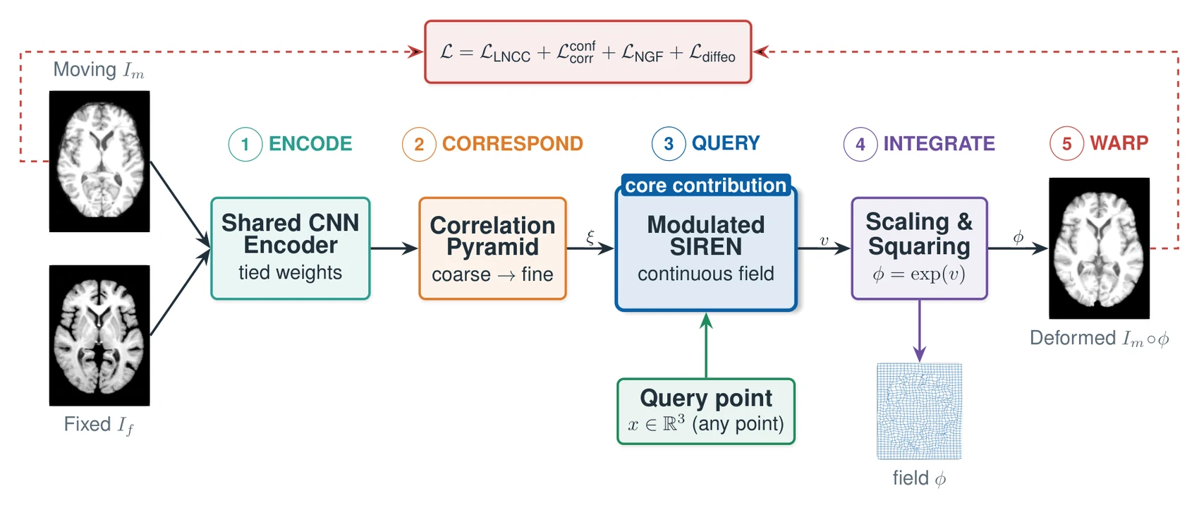
A modulated SIREN decodes the velocity field as a continuous function of coordinates, over a coarse to fine correlation pyramid. Scaling and squaring integrates that velocity into the deformation. The whole model is 5.51 million parameters and trains without ever seeing a segmentation label.

Encode, correspond, query, integrate, warp. The SIREN in the middle is the part that makes the field a function rather than an array.
4 items. Drag, scroll, or use the arrows.
Where this goes next
The continuous query in QueryPhi concerns the decoder output. The encoder and the correlation pyramid still run at the resolution the model was trained on, so the paper is explicit that robustness to different input voxel spacings, and to anisotropic acquisition, is not something it demonstrates.
That gap is the most interesting thing about the result. A continuous field should earn its keep exactly where sampling is the binding constraint, and on 1 mm isotropic brain MRI sampling is generous. Most clinical MRI outside neuroimaging is not: an abdominal sequence might be 1.5 mm in plane and 7 mm between slices, close to five to one.
So the work in progress is a controlled study on a benchmark that contains both, with anisotropy as a dial inside one dataset rather than a difference between datasets, the endpoint pre-specified as topology rather than accuracy, and every method held to the same protocol. Early search level runs are encouraging and are nowhere near the fidelity needed to make the claim, which is why the paper lists this as a limitation rather than a finding.

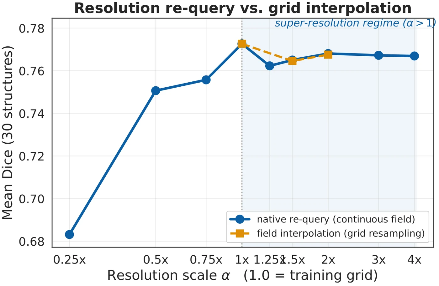
The field can be re-queried at any output resolution once trained. Whether that also holds for anisotropic input sampling is the open question, and the paper says so rather than implying otherwise.
CVPR 2026, PVUW Workshop · co-author
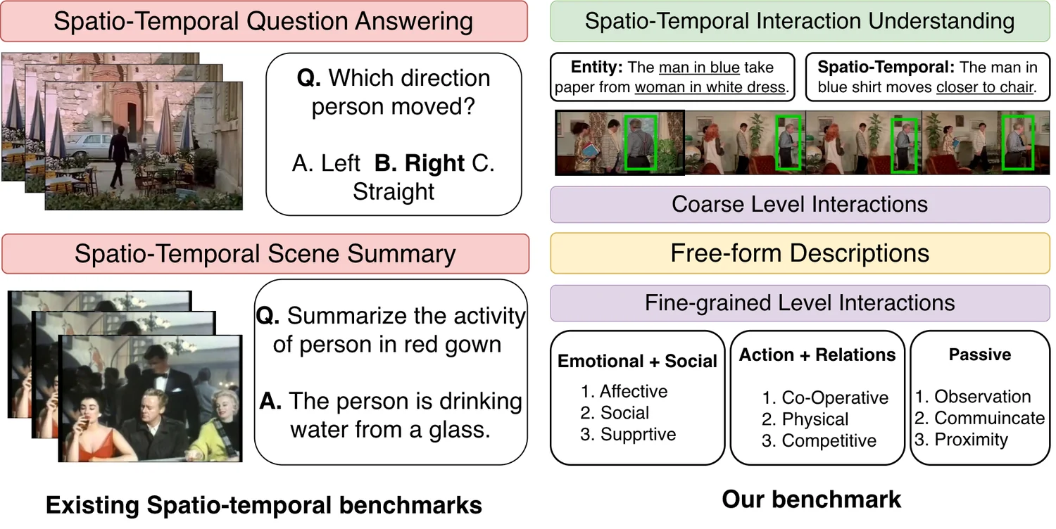
A benchmark asking whether vision language models actually understand interactions in video, or recognise objects and guess the rest.

Across 11 state of the art models, breaking aggregate performance down along the taxonomy revealed pronounced spatio-temporal biases that traditional metrics obscure completely. Models that looked comparable in aggregate turned out to behave very differently once the axes were separated.
VISTA is the first large scale interaction aware diagnostic benchmark for spatio-temporal understanding in vision language models.
Projects
Course work, challenge entries and implementations written from scratch. Each one is here because it taught me something that shows up in the work above.

Four training runs on paediatric brain tumor MRI, held identical except for the loss and the optimiser, then read through the curvature of the loss surface rather than the leaderboard.
Writing
Longer pieces on how the method came together, what the measurements said, and the experimental habits I picked up on the way.
How I got here
I went toward brain imaging for reasons that were more philosophical than technical, and the interest moved once I arrived. What holds me now is anatomy, and the geometry and optimisation sitting underneath the methods that measure it.
The habit I cannot put down is wanting to know which part of a result is doing the work. Most of what I have built since is an attempt to make that question answerable rather than arguable.
Hover for colour, click to enlarge.
Currently
University of Central Florida
AI MIND Lab, University of Central Florida
Center for Research in Computer Vision, UCF
Medical imaging, continuous and physically constrained representations, uncertainty, and reconstruction from incomplete measurements. Happy to share the manuscript.